
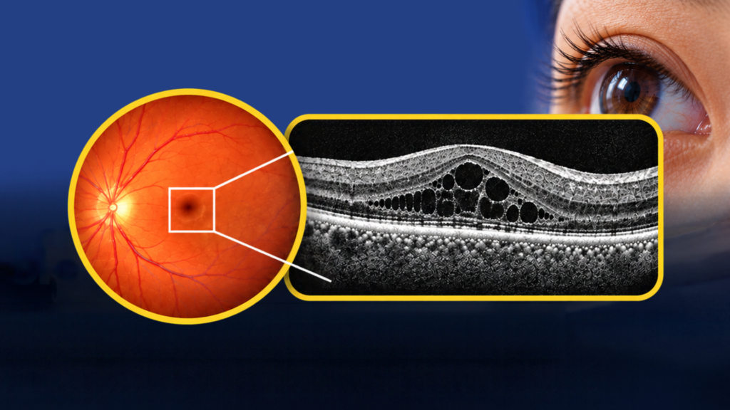
For people with diabetes, clear vision doesn’t always mean the retina is healthy. Over time, high blood sugar levels can damage the tiny blood vessels at the back of your eyes, allowing fluid to leak into the surrounding retinal tissue. When this leakage causes swelling in the macula, the part of your retina responsible for sharp, detailed vision, it is known as diabetic macular oedema (DME).
The surprising part? Your retina can already be swollen, even when your vision seems perfectly normal. Some people experience blurry or distorted vision, while others may notice no changes at all, especially when the swelling is mild or does not affect the centre of the macula.
So, how can we detect these hidden changes before they start affecting your vision? That’s where Optical Coherence Tomography (OCT) comes in. This advanced eye imaging technology allows us to examine the retina layer by layer, helping reveal swelling and structural changes that may otherwise go unnoticed.
At the back of your eye is a thin, light-sensitive layer called the retina. Think of it like the sensor in a camera. It receives light and sends visual information to your brain, helping you see the world around you. Near its centre is the macula, which allows you to see fine details when reading, recognising faces or using your phone.
When fluid leaks into the retinal tissue, it can accumulate within the retinal layers, causing them to become thicker and swollen. Think of a sponge absorbing water. As it absorbs more fluid, it becomes thicker and expands. Something similar can happen to your retina.
If swelling affects the macula, particularly its centre, it can interfere with your ability to see fine details clearly. However, the amount of swelling and how well you can see do not always go hand in hand. This is why relying on clear vision alone is not enough to assess your retinal health.
Imagine looking at a cake. From the top, you can only see its surface. But when you cut a slice, you can see the different layers inside. OCT works in a similar way, except it uses light to scan your retina without touching your eye.
During a retinal examination, our optometrists can look for visible signs of diabetic eye disease. However, some changes within the retinal layers may be difficult to identify through a conventional retinal examination alone. OCT provides detailed cross-sectional images, allowing us to examine the retina layer by layer.
With OCT, we can look for:
Retinal thickening: Areas where the retina has become thicker due to swelling.
Fluid-filled spaces: Small pockets of fluid within the retinal layers.
Changes in retinal structure: Irregularities that may be associated with swelling or other retinal conditions.
OCT also measures retinal thickness, helping us assess the extent of swelling and monitor changes over time. In some cases, OCT can identify diabetic macular swelling before noticeable changes in vision develop. This makes it a valuable tool for detecting and assessing suspected diabetic macular oedema, particularly when subtle retinal changes are not easily visible during a conventional examination.
Although OCT provides detailed images of the retinal layers, it is only one part of assessing diabetic eye health. OCT helps detect retinal swelling, but it does not replace a comprehensive diabetic retinal examination.
Diabetes can also cause other retinal changes, including tiny haemorrhages, abnormal blood vessels and areas of reduced blood supply, which may not be fully assessed through a standard OCT scan alone. A comprehensive diabetic retinal examination helps identify signs of these changes and evaluate your overall retinal health.
If diabetic macular oedema is detected or suspected, prompt referral to an ophthalmologist experienced in diabetic retinal disease is important for further evaluation and appropriate management.
For adults with type 2 diabetes, an initial comprehensive retinal examination is recommended at diagnosis. For adults with type 1 diabetes, the Malaysian Ministry of Health’s diabetic retinopathy screening module recommends initial screening within three years after diagnosis.
The recommended follow-up interval depends on whether diabetic retinal changes are present and other risk factors. People with existing retinopathy may require more frequent monitoring, while those with consistently normal findings may be advised to return at longer intervals depending on their condition.
Your optometrist or eye care professional can advise on an appropriate examination schedule based on your individual eye health and medical history.

Your vision may still be clear, but changes could already be happening inside your retina. Regular diabetic retinal examinations help identify changes early, while OCT provides a closer look at the retinal layers when further assessment is needed.
At Eye & Glasses Optometrist, our optometrists can assess your retinal health and advise whether an OCT scan is appropriate as part of your examination.
This November, in conjunction with Diabetic Eye Disease Awareness Month, take a proactive step towards protecting your vision. Don’t wait until your vision becomes blurry to find out what’s happening inside your eyes.
Book an Advanced Eye Examination with Eye & Glasses Optometrist to better understand your retinal health.